
When your pet has been scheduled for an ultrasound examination, we understand that it’s helpful if you know what to expect. Just like with people, the purpose of this procedure is to aid in making a proper diagnosis of a disease-causing illness or other condition. Ultrasound can give your dog or cat’s veterinarian so much information about what is happening on the inside - in real time. But what are we looking for, how should you prepare your pet, and what will the results tell us?
Let’s look at some frequently asked questions about pet ultrasound.
What is an Ultrasound Machine?
An ultrasound machine emits ultrasound waves that penetrate into your pet’s organs. These waves are reflected back into the hand-held probe that is placed on the skin. The pattern of the 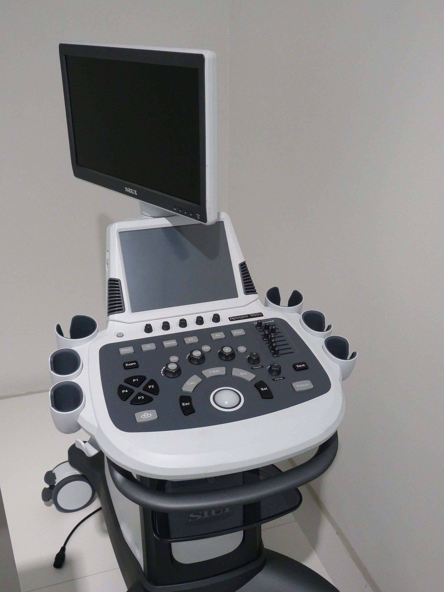 reflected sound waves creates an image that is viewed on a screen.
Is Radiation Involved?
Unlike X-rays, radiation is not part of an ultrasound examination. As the name suggests, this type of imaging works via sound waves. That means that your dog or cat, plus the veterinarian and staff, would not be exposed to any radiation from proximity to an ultrasound machine.
Which Types of Disease are Typically Diagnosed with an Ultrasound Examination?
The ultrasound examination permits a detailed view of many of the body’s organs that may not be available with other diagnostic imaging methods. For example, the kidneys can be seen on X-rays, but only their size and shape can be determined, whereas ultrasound allows us to actually view the internal structures of these and other vital organs.
An ultrasound examination is especially helpful for diseases of the heart. An ultrasound of the heart is called an echocardiogram, or “echo.” Via ultrasound, the heart’s wall thicknesses and the size of its chambers can be determined. Visualization of the valves determines whether they are functioning properly. Motion can also be detected, which gives us an assessment of the heart's ability to move blood.
Some diseases can be diagnosed with this method because they have a specific ultrasound appearance. Others, however, produce ultrasound findings that are not definitive. In these cases, we would choose another imaging method, or even a combination of methods, to gather the diagnostic information needed to provide the best care for your dog or cat.
What Could Be the Next Steps After an Ultrasound?
One of the important features of an ultrasound examination is the ability to find abnormal areas in the organs. This permits precise biopsy of those areas, particularly because the live, moving ultrasound image can serve as a guide for a veterinarian to obtain a sample of the area in question. This can be done, for example, by inserting a needle that can be seen on the ultrasound image and obtaining cells from the organ, or even a sterile urine sample from the bladder.

A technician may test a sample, or a pathologist could examine the biopsied section of tissue under a microscope to gain more information. In many cases, the ultimate diagnosis is made by the pathologist.
What Steps Need to be Taken to Prepare for an Ultrasound Exam?
Special preparation would likely not be necessary for an echocardiogram. However, if organs in the abdomen are to be studied, we may instruct you to withhold food from your pet for the 12 hours ahead of the exam.
Additionally, the urinary bladder is best visualized if it is full of urine. Therefore, your pet should not urinate within three to six hours of the study, if possible. Cover or put away litter boxes for cats before you head to your appointment. For smaller dogs, you can try to keep them from urinating by carrying them into our office. For larger dogs who will walk on-leash and will want to explore and “mark” while they walk, try to encourage them to move quickly. Get them excited, maybe even by jogging, through our front door, so they don’t have time to empty their bladder anywhere. If you need any other tips for keeping your pet’s bladder full for an ultrasound, please get in touch!
If anesthesia is needed, there may be other steps to prepare your pet for the ultrasound procedure.
Is Anesthesia Required?
If your pet is calm and/or cooperative, no anesthesia or sedation is needed to perform an ultrasound on the heart or the abdomen. However, if biopsies are to be taken, a short-acting anesthetic will be needed to help prevent complications.

We may ask you to withhold food and water ahead of your pet’s visit, should anesthesia be required. You will receive specific instructions for your dog or cat if this is the case.
Is it Necessary to Shave My Pet's Hair?
In most cases, yes. It is important that the hand-held ultrasound probe makes complete contact with the skin. Sometimes the hair can be moistened with alcohol, but most studies require hair removal. Our staff will take care of this step before the exam, and we’ll work to keep any “haircuts” as neat and even as possible!
Which Organs Cannot Be Studied with Ultrasound?
Air is the enemy of ultrasound waves. Since the lungs are filled with air, they cannot be studied. An exception is made for a mass that is located within the lungs. Bone also stops ultrasound waves, so the brain and spinal cord are not seen with an ultrasound study. The skeletal system itself is also not examined with ultrasound.
When Will I Know the Results of the Examination?
Since an ultrasound study is performed in real time, the visual results are known immediately. In some cases, the ultrasound images are sent to a veterinary radiologist for further consultation. If this happens, the final report may not be available for a few days. We will let you know what to expect once the ultrasound exam is complete.
If you have any other questions about ultrasound and veterinary digital imaging, please reach out to us! We’re happy to talk with you about your dog or cat’s health care needs.